Diagnostic imaging includes all tests that produce images or pictures of the inside of the body in order to diagnose diseases. These tests may use x-rays, sound waves, radio waves, and radioactive waves and particles that are recorded by photographic films or other types of detectors.
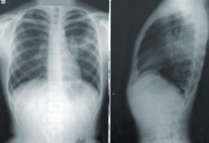
General Radiography
Examination of any part of the body for diagnostic purposes by means of x-rays with the record of the findings usually impressed upon a photographic film.
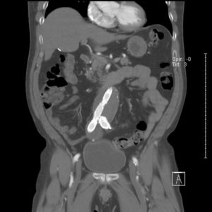
CT (“Cat Scan”)
Computed tomography (CT) is a noninvasive medical test that helps physicians diagnose and treat medical conditions. CT imaging combines special x-ray equipment with computers to produce multiple images or pictures of the inside of the body. These cross-sectional images of the area being studied can then be examined on a computer monitor or printed.
Fluoroscopy
Fluoroscopy is examination of the tissues and deep structures of the body by x-ray, using a fluoroscope. The fluoroscope is a device that projects radiographic (x-ray) images in a movie-like sequence onto a screen monitor. Common fluoroscopic studies include barium swallow and upper GI to evaluate the function of the esophagus and stomach, barium enema to evaluate the large intestine, and retrograde urethrograms to evaluate the urinary system.
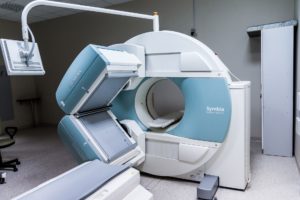
MRI (Magnetic Resonance Imaging)
Magnetic Resonance Imaging is a noninvasive medical test that helps physicians diagnose and treat medical conditions. MR imaging uses a powerful magnetic field, radio frequency pulses and a computer to produce detailed pictures of the internal body structures. The images can then be examined on a computer monitor, printed or copied to CD. MRI does not use ionizing radiation (x-rays).
Ultrasound
Ultrasound imaging is a noninvasive medical test that helps physicians diagnose and treat medical conditions. Ultrasound imaging, also called ultrasound scanning or sonography, involves exposing part of the body to high-frequency sound waves to produce pictures of the inside of the body. Ultrasound exams do not use ionizing radiation (as used in x-rays ). Because ultrasound images are captured in real-time, they can show the structure and movement of the body’s internal organs, as well as blood flowing through blood vessels.
Nuclear medicine
Nuclear medicine uses radioactive substances to diagnose and treat disease. A variety of different radioactive substances may be ingested or injected into the body. A special camera then detects the activity produced by these substances to create images which are then interpreted by the radiologist. Types of nuclear medicine tests include PET scans and bone scans for cancer surveillance, cardiac perfusion scans to evaluate the function of the heart, thyroid scans for both diagnosis and treatment of thyroid diseases, HIDA scans to evaluate the gallbladder, and gastric emptying studies to evaluate the function of the stomach.