Breast Imaging

Athens Radiology Associates offer the full complement of state-of-the-art breast imaging services. We have six subspecialized breast imaging physicians and a team of nurses, technologists, and sonographers all with a passion for excellent and compassionate care.

Screening Mammogram

The goal of breast cancer screening is to reduce deaths due to breast cancer by detecting cancer at an early stage when treatment is more effective. For most women, yearly mammograms should begin at age 40. Most women who are diagnosed with breast cancer do not have a family history, and 1 in 8 women will be diagnosed with breast cancer in their lifetime. Multiple studies have shown that annual screening mammograms starting at age 40 saves the most lives. See below to learn more about breast cancer screening and how we evaluate breast cancer risk.

Breast cancer screening

• According to National Cancer Institute data, since screening mammography became widespread in the early 1990’s, the U.S. breast cancer death rate has decreased by over 30%.

• Mammography is not perfect, and it will not benefit all women equally. Mammography detects most, but not all, breast cancers. Monthly self breast exam and a yearly clinical breast exam by your doctor are also important in detecting any suspicious changes in your breasts.

• Major health organizations including the American College of Obstetricians and Gynecologists, the American Cancer Society, the American Society of Breast Disease, the American College of Radiology, and the Society of Breast Imaging agree that starting annual mammography at age 40 saves the most lives.

• For some women at higher risk for breast cancer, yearly screening mammograms should begin at age 30, and yearly screening MRI can also begin as early as age 25.

• For more information about screening mammography, visit the Society of Breast Imaging Patient Resources page: https://www.sbi-online.org/endtheconfusion/PatientResources.aspx

Lifetime risk for breast cancer

• Lifetime risk for breast cancer is calculated based on a combination of personal history and family history factors. Multiple models for calculating breast cancer risk exist. At our breast centers, we use the Tyrer-Cuzick v8 model, which is considered one of the most comprehensive models and also takes breast density into account.

• Average lifetime risk for breast cancer in the United States is 11%. Women who are at 15-19% lifetime risk for breast cancer are considered intermediate risk. Women who are at 20% or greater lifetime risk for breast cancer are considered high risk.

• You may be at increased lifetime risk for breast cancer if you have any of the following:

– A family history of breast cancer

– A personal history of breast cancer or breast atypia

– A family or personal history of a genetic mutation known to predispose to breast cancer. These include but are not limited to BRCA1, BRCA2, ATM, P53, CHEK2, PTEN, CDH1, STK11, PALB2.

– Ashkenazi Jewish descent

• Additional factors that influence risk for breast cancer include age of your first period, age when you had your first child, age of menopause, weight, use of hormone replacement therapy, and breast density.

• Talk to your doctor to assess your lifetime risk for breast cancer and discuss which screening tests are right for you. A free, online risk assessment model is available here: https://ibis.ikonopedia.com/ (Please note that this model may provide a slightly different result than the model used by your doctor or by our breast center).

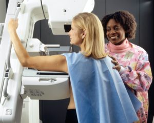
Diagnostic Mammogram

A diagnostic mammogram is performed for women who are having breast symptoms such as a lump, focal pain, nipple discharge, or skin changes. It is also performed for women who have had an abnormality detected on their screening mammogram. A diagnostic mammogram involves additional dedicated views to closely examine an area of interest in the breast. A breast imaging radiologist will review your images right after they are taken to determine whether additional images or a breast ultrasound is needed. You will be given your results by the end of your appointment.

More info

• About 1 in 10 women will be called back for additional imaging after their screening mammogram.

• Most of the time, the additional imaging will show no evidence of cancer and you will be returned to screening mammogram. Sometimes, a follow up exam in a few months will be recommended.

• Sometimes, a needle biopsy will be needed to determine whether a spot in the breast is cancer. Biopsies are performed by our dedicated breast imaging radiologists who have expertise in image guided biopsies. (See Biopsy section below).

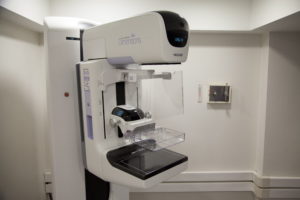
3D-Mammography (Digital Breast Tomosynthesis)

Digital breast tomosynthesis (tomo), also known as 3D mammography, is a screening and diagnostic breast imaging tool to improve the early detection of breast cancer. During the 3D part of the exam, an x-ray arm sweeps over the breast, taking multiple images in seconds that can then be viewed by our radiologists. 3D mammography increases cancer detection rate and decreases callback rates from screening and is particularly helpful to evaluate mammograms for women with dense breast tissue. See below to learn more about 3D mammography and breast density.
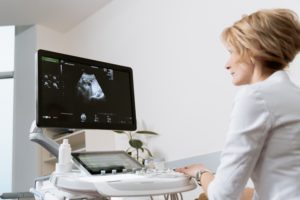
3D Mammography

• Approved as an imaging modality by the FDA in early 2011, 3D mammography is now routinely used at our breast imaging centers.

• 3D mammography increases cancer detection rate and decreases callback rates from screening. • 3D mammography is particularly helpful for women with dense breast tissue because it allows the radiologist to better see through the layers dense tissue.

• 3D mammography does involve slightly higher radiation exposure than 2D mammography, but the overall radiation dose is still very low (significantly less than a CT scan). The radiation dose for a combined 2D/3D mammography exam is well below the acceptable limits defined by the FDA, and there is no evidence that this low level of radiation has any harmful effect on the breasts.

• 3D mammogram is covered by most insurance companies and Medicare. You do not need a separate order from your doctor other than the standard screening or diagnostic mammogram order. If your insurance does not cover the 3D mammogram there will be an additional charge that you will be responsible for in addition to the fee to the radiologist to interpret the exam. You may be able to pay this with your flexible spending account.

Breast Density

• Breast Density is an assessment of the amount of fibrous and glandular tissue compared with the amount of fatty tissue in the breast. (Dense breast tissue appears white on a mammogram and fatty tissue appears black). This is important because of “masking effect” which means denser breast tissue can hide cancer.

• About half of women have non-dense breasts. The other half of women have dense breasts.

• There is a small increase in breast cancer risk for women with dense breast tissue. This increase is usually not enough to put you in a higher lifetime risk category without additional risk factors for breast cancer. (See Lifetime risk for breast cancer section above).


Breast Ultrasound

Ultrasound is a noninvasive medical test that uses sound waves to produce pictures of the internal structures of the body. A breast ultrasound may be performed as part of your diagnostic exam. You will be scanned by a sonographer as well as by a specialized breast imaging radiologist. The radiologist will go over the results and recommendations with you at the end of your exam.

Breast MRI

Magnetic resonance imaging (MRI) is a noninvasive medical test that uses a powerful magnetic field without radiation to produce pictures of the body which are then examined on a computer monitor. ARA’s highly trained imaging specialists use state of the art equipment and software to produce and interpret breast MRI images.

More info

• Breast MRI can be used to screen women at high risk for breast cancer. In the screening setting it is used in conjunction with screening mammography. It is not a replacement for mammography.

• MRI of the breast can be used as a tool for pre-surgical evaluation of patients with known breast cancer, and as a problem-solving tool when mammography and ultrasound do not provide clear answers.

• Breast MRI is very sensitive for detecting breast cancer. However, it can sometimes be too sensitive and also pick up spots in the breast which are not cancer. For this reason, it is not recommended for everyone. Talk to your doctor to determine if a breast MRI is recommended for you.

Image-Guided Breast Biopsy

Lumps or abnormalities in the breast are often detected by physical examination, mammography, or other imaging studies. However, it is not always possible to tell from these imaging tests whether a lump is benign or cancer. A core needle breast biopsy is performed by a highly trained radiologist to remove some cells through a hollow needle from a suspicious area in the breast and examine them under a microscope to determine a diagnosis.

More info

• ARA breast imaging radiologists are highly trained in performing image guided needle biopsies using multiple imaging modalities including ultrasound, stereotactic or tomosynthesis (both using a mammogram machine), and MRI.

• These biopsies are performed at our breast imaging centers. They are minimally invasive (not surgery) and use local anesthesia to numb the breast tissue.

• If you do need a breast biopsy, a dedicated nurse navigator will guide you through the entire process from scheduling the appointment to discussing your results and setting up any follow up appointments you may need. After the pathology results are available, a breast radiologist will review your imaging to make sure that the pathology results match what we see on the imaging. Based on the result, the radiologist will make a recommendation for follow up imaging or surgical consultation if needed.

Breast Needle Localizations

For women who need breast surgery to remove a cancer or atypical lesion, we can perform an image guided localization to help the surgeon know exactly which area to take out.  Ultrasound or mammography is used as a guide to put a wire or radiofrequency reflector through the abnormality in the breast. During the surgery, the surgeon uses the wire or reflector to locate and remove the abnormality. Since most breast cancers we diagnose by mammography are too small to feel, performing an image guided localization helps the surgeon remove the entire abnormal area while conserving as much normal breast tissue as possible.

Ultrasound Guided Cyst Aspiration

Breast cysts are benign sacs of water that form in the breast. They are very common and do not cause a problem for the majority of women. For some women, a cyst may become large and painful. In these situations, we can drain the fluid from the cyst using a small needle under ultrasound guidance.

Consultations for review of outside exams

ARA’s radiologists can review studies you have had done elsewhere and give a second opinion.

Billing Info

Payment System

When your services are provided in a hospital setting, you will receive an invoice from Athens Radiology Associates, P.C. for the interpretation of your images. You will also receive a separate invoice from the hospital for the technical fee related to generating and creating your images.


For account and patient billing information, please call:
855-826-6459


Payments can be mailed to:

Athens Radiology Associates
PO Box 106062
Atlanta, GA 30348

Radiology Associates of North Georgia
PO Box 161246
ATL 30321

Contact Us

PO Box 7879
Athens, GA 30604

Office Phone: 706-850-5001
Fax: 706-521-8593
  • This field is for validation purposes and should be left unchanged.