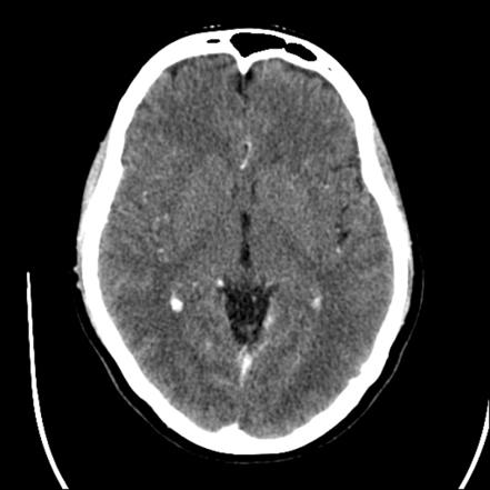

CT of the head, neck, and spine
CT scanning, sometimes called “CAT scanning”, is a noninvasive medical test that helps physicians diagnose and treat medical conditions. CT imaging combines special x-ray equipment with sophisticated computers to produce multiple images or pictures of the inside of the body. These cross-sectional images of the area being studied can then be examined on a computer monitor or printed.
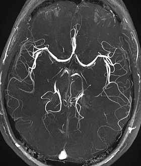
MR and CT Angiography of the brain and neck
Angiography is a minimally invasive medical test that helps physicians diagnose and treat medical conditions. In magnetic resonance (MR) angiography, a powerful magnetic field, radio waves and a computer produce the detailed images. MR angiography may be performed with or without contrast material. In CT angiography, special x-ray equipment produces cross-sectional images after contrast is injected. If needed, the contrast material is usually injected using a vein in the arm.
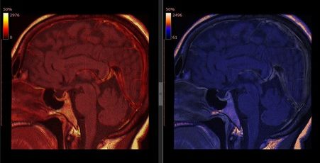
Ultrasound
Ultrasound of the brain, spine, neck, and thyroid.
Ultrasound and CT guided biopsies of the head, neck, spine.
Myelograms
In this procedure, contrast dye is injected into the spinal canal, allowing spinal cord and nerve compression caused by herniated discs or fractures to be seen on an x-ray.
Fluoroscopy-guided lumbar punctures
Fluoroscopy is a study of moving body structures. It’s similar to an x-ray “movie.” A continuous x-ray beam is passed through the body part being examined, and is transmitted to a TV-like monitor so that the body part and its motion can be seen in detail.
Discography
Involves the injection of a special contrast dye into a spinal disc thought to be causing low back pain. The dye outlines the damaged areas on x-rays taken following the injection.